
Ideaquest Technology
Column: FG vision sensor
Medical and social welfare systems that we are developing are based on technology which detects minute body movements without contacts or constraints. The FG vision sensor is the key component of our system. The FG vision sensor consists of a semiconductor laser, FG (Fiber Grating) element and a CCD camera. When a laser beam is projected onto the 5mm square FG element, the beam is split into 2000 spots by diffraction. These spots are captured by the CCD camera and the movements of the spots are calculated in order to determine the minute movements of the body without any contacts or any invasion.
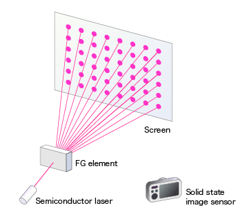
Figure 1 Construction of FG vision sensor
If the screen is flat, the light spots line up regularly. On the other hand, if the screen is uneven, the light spots are shifted according to the heights of the unevenness as per Figure 2. Figure 3 depicts the image captured by the CCD camera in such cases. If the light spots are shifted to the right, the screen is protruded. If the light spots are shifted to the left, the screen is caved in.
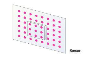
Figure 2 Uneven screen
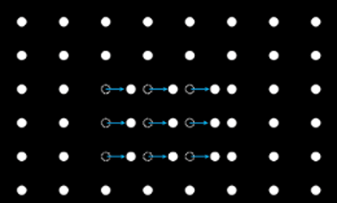
Figure 3 Image from the solid state image sensor
Respiration makes the chest move. Swallowing food makes the surface of the throat move. By capturing the light spots on the chest or throat and by analyzing the motion of the spots, minute motion of the body can be detected in chronological order. Figure 4 depicts respiration waveforms acquired by accumulating the temporal movement.

Figure 4 Acquisition of respiration waveforms
On the other hand, by plotting the back and forth movements of light spots on the throat, the 3 dimensional shape of the throat surface can be captured. By analyzing the temporal change of the throat surface the swallowing capability can be evaluated.
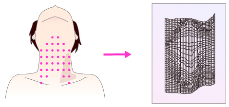
Figure 5 Acquisition of 3 dimensional image from the light spots on throat
Thus, we developed non-contact and non-invasive analysis and monitoring systems using FG vision sensor.
Moreover, a filter which only passes the projected light is placed in front of the solid state image sensor. Thus, only the light spots are recorded by the camera. The image of the patient is not recorded. The FG vision sensor protects the privacy of the patient and only record necessary information.